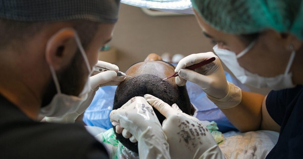
A man wrote to me last spring. He was 34, had been losing hair from the crown for about two years, and had been on finasteride and minoxidil for eighteen months. He had switched to topical finasteride six months in, then added microneedling, then added oral biotin, then added a peptide serum from Instagram. The hair was still going. He sent me a photo of his scalp under good light and the diagnosis was on the photo. It was not androgenetic alopecia. It was follicular tufting with peripheral pustules. He had folliculitis decalvans. He had spent eighteen months treating the wrong condition while a scarring alopecia ate his follicles.
This is the most consequential diagnostic miss in scalp medicine and it happens constantly. The two conditions look similar from across a room. They look different on close examination. The treatments are nearly opposite. The window to intervene on the scarring version is narrow.
What the studies actually show
Androgenetic alopecia is the common pattern hair loss most people picture when they think of hair loss. It is mediated by dihydrotestosterone acting on follicular androgen receptors, miniaturizing terminal hairs into vellus hairs over years to decades. The follicle does not die, it shrinks. This means that an aggressive intervention at the right time can restore terminal hair. Olsen 1999 in the JAAD and subsequent finasteride trials established the diagnostic criteria and the reversibility window.
Folliculitis decalvans is a primary cicatricial alopecia. It is an inflammatory condition that destroys the follicle and replaces it with scar tissue. Once a follicle is gone it is gone. The pattern is patchy hair loss with peripheral pustules, follicular tufting where multiple hairs emerge from a single dilated ostium, perifollicular erythema, and crusting. Trüeb and colleagues in their 2018 trichology research in International Journal of Trichology (PMID: 30574255) catalogued the clinical and dermoscopic features and emphasized how often the condition is misread as common androgenetic alopecia or as ordinary folliculitis that should respond to over-the-counter shampoos.
The 2020 JAAD review by Vano-Galvan and colleagues on cicatricial alopecias (PMID: 32531385) reported that the average time from first symptom to correct diagnosis of folliculitis decalvans is 2 to 4 years. During those years the inflammation destroys follicles that will never grow back. The diagnostic delay is the disease.
Dermoscopy distinguishes them clearly when you know what to look for. Androgenetic alopecia on dermoscopy shows hair shaft diameter variability of more than 20 percent, peripilar brown halos, and yellow dots in advanced cases. Folliculitis decalvans shows follicular tufting (six or more hairs from one ostium), perifollicular hyperkeratosis with yellow-tubular scales, perifollicular erythema, and pustules. These are visible on a 10x dermoscope. They are not visible on Instagram before-and-afters. They are not visible to a barber who is trying to help.
The contrarian section
The wellness industry around hair loss has built a vocabulary that does not distinguish between these conditions. “Hair regrowth” is treated as one category. Rosemary oil, peppermint oil, scalp serums, microneedling, derma stamps, red light caps, biotin, collagen powder, the entire content economy of male and female hair loss is sold on the assumption that hair loss is one thing and intervention is gradient.
This is wrong in a way that has cost real follicles. Microneedling on an androgenetic alopecia scalp is a reasonable adjunct with modest data, per the Dhurat 2013 trial in the Journal of Cutaneous and Aesthetic Surgery. Microneedling on a folliculitis decalvans scalp is potentially harmful because mechanical trauma can extend the inflammatory process and accelerate follicular destruction. The same intervention helps one condition and worsens the other. The Instagram content does not distinguish.
Topical finasteride for androgenetic alopecia is well-supported. Topical finasteride for what looks like androgenetic alopecia but is actually a scarring inflammatory condition does nothing for the inflammation and lets the follicles die on schedule. The patient who wrote to me had been told by three different telemedicine prescribers that his pattern was male pattern baldness. None of them looked at his scalp under magnification. None of them asked about pustules. None of them asked about itching or burning, which are common in folliculitis decalvans and absent in androgenetic alopecia.
The signs that should trigger reassessment are specific. Itching or burning of the scalp in the area of hair loss. Pustules at the hair line of the bald patch. Crusting or yellow scaling that is not seborrheic dermatitis. Hair coming out in clumps when the patches are pulled. Multiple hairs emerging from one follicle, visible on close inspection or photography. Patchy rather than symmetric pattern. Sudden acceleration of loss rather than slow progression. Any of these should send a patient to a dermatologist with a dermoscope, not back to the finasteride prescription.
The treatment of folliculitis decalvans is anti-inflammatory and antimicrobial. The standard protocol per the 2018 trichology literature is combined topical and oral antibiotics, often a tetracycline plus topical clindamycin, sometimes adding hydroxychloroquine or short-course systemic steroids for refractory cases. Isotretinoin has been used. The goal is suppression of the inflammatory cascade before more follicles are destroyed. This is not a goal that minoxidil or finasteride addresses.
What I would tell my past self
If I were thinking about hair loss in a male or female patient at any age, I would not start with the assumption of pattern baldness. I would start with a magnified photograph of the affected area in good light. I would look for the specific features that distinguish androgenetic alopecia from cicatricial conditions. I would ask about symptoms beyond just visual loss. Itching. Burning. Crusting. Tenderness on touch. Any of these would change the differential.
I would also resist the telemedicine flow that prescribes finasteride based on a smartphone photo and a checkbox form. The form does not ask the right questions because the form is designed for the high-prevalence condition. Folliculitis decalvans is rare enough that the screening flow misses it, and common enough that several thousand people a year in the US alone get the wrong treatment. The economic incentive of the telemedicine model is volume. The diagnostic precision required for cicatricial alopecia is not in the model.
If you have noticed your hair thinning, I would have a dermatologist who specializes in hair, called a trichologist in some practices, look at your scalp under magnification before starting any treatment. The 30 minutes of consultation cost might save you years of follicles. Once a scarring alopecia has destroyed a follicle there is no rescue. Hair transplant into scarred scalp has a poor take rate because the recipient tissue is fibrosed. Prevention is the only window.
I would also have warned myself about the supplement-and-serum content economy. Almost none of it distinguishes between conditions. Biotin does nothing for either androgenetic alopecia or cicatricial alopecia. Collagen powder does nothing. Peptide serums for hair loss are mostly minoxidil analogs without the minoxidil. Rosemary oil has one positive trial against minoxidil in androgenetic alopecia (Panahi 2015) and that trial is the only piece of evidence supporting an entire content category.
The thing I would say plainly is that hair loss is a medical condition and the right specialist for it is a dermatologist trained in trichology. Not a stylist, not a telemedicine intake form, not a TikTok hair coach. The cost of misdiagnosis is permanent. The benefit of correct diagnosis is sometimes also permanent.
Frequently asked
How common is folliculitis decalvans compared to androgenetic alopecia?
Folliculitis decalvans accounts for roughly 11 percent of primary cicatricial alopecias seen in specialist trichology clinics, per the 2020 JAAD review. Cicatricial alopecias as a category are about 7 percent of all hair loss diagnoses in dermatology. Androgenetic alopecia is the majority. But the absolute numbers of folliculitis decalvans patients are large enough that any dermatologist sees several per year, and any telemedicine prescriber writing finasteride scripts is statistically encountering them and not catching them.
Can folliculitis decalvans be cured?
Cured in the strict sense, no. Suppressed and stabilized, yes, in most cases. The standard antibiotic protocols achieve clinical quiescence in roughly 60 to 80 percent of patients with adequate adherence and duration. The lost follicles do not return but the active inflammation can be controlled and progression halted. Long-term maintenance therapy is often required.
What does dermoscopy of androgenetic alopecia look like that distinguishes it?
Hair shaft diameter variability greater than 20 percent within a single field is the most reliable sign of androgenetic alopecia. This means you can see thick terminal hairs next to thinner intermediate hairs next to vellus hairs in the same area. Folliculitis decalvans does not show this miniaturization gradient because the destruction is binary. The follicle is either functioning or scarred over.
Should I get a scalp biopsy?
A scalp biopsy is the gold standard for cicatricial alopecia diagnosis. A 4 mm punch biopsy at the active edge of a lesion, processed for both vertical and horizontal sections, can distinguish folliculitis decalvans from lichen planopilaris from frontal fibrosing alopecia from central centrifugal cicatricial alopecia. If your dermatologist is unsure on dermoscopy and the pattern is atypical for androgenetic alopecia, a biopsy is worth doing.
What about lichen planopilaris and frontal fibrosing alopecia? Are those also missed?
Yes, and often more so than folliculitis decalvans. Frontal fibrosing alopecia in particular has become much more common in the last 20 years and is frequently missed because the hairline recession looks like androgenetic alopecia in older women. The clue is loss of eyebrow hair, loss of body hair in unusual places, and perifollicular erythema at the hairline. If you are an older woman with progressive hairline recession and your eyebrows have thinned, see a trichologist.
Sources: Trüeb et al. Int J Trichology 2018, PMID: 30574255. Vano-Galvan et al. JAAD 2020, PMID: 32531385. Olsen 1999 androgenetic alopecia diagnostic criteria. Dhurat et al. 2013 microneedling for androgenetic alopecia. Panahi et al. 2015 rosemary oil trial.
Related Elelaf tools
- Scalp bumps decoder
- Hair growth protocol
- Scalp flakes
- Face redness reset
- Sebderm rosacea eczema decoder
References
- Kligman AM, Christensen MS. The biology of the stratum corneum revisited. Int J Cosmet Sci. 2011. PubMed.
- Draelos ZD. The science behind skin care: cleansers. J Cosmet Dermatol. 2008. PubMed.
Have a question about “Folliculitis decalvans vs androgenetic alopecia: the scalp diagnosis most patients miss”?
Ask our editorial desk. Best questions become full follow-up articles, reviewed by our medical reviewer. No medical advice given in private — answers run as articles or not at all.